
The amniotic membrane is the layer that covers the inner surface of the placenta. When this temporary organ is expelled after birth, it becomes biological waste. Its tissues, when processed and preserved, can be reused for medicinal purposes thanks to their proven anti-inflammatory and regenerative properties. This is why placental material is widely used worldwide for various tissue repair treatments, with applications ranging from eye and dental injuries to complex skin wounds.
In an CONICET’s article signed by Marcelo Gisande we can read that recently, a CONICET research team, comprised of professionals from the Institute of Immunological and Physiopathological Studies (IIFP, CONICET-UNLP-associated with CICPBA) and Unit 4 of the Center for Translational Medicine (U4-CEMET, HEC), helped verify the effectiveness of amniotic membrane-derived patches or dressings for treating complex wounds. They achieved complete closure of a recalcitrant ulcer and successful tissue regeneration in an oncology patient undergoing surgery. The results of the procedure were published in the scientific journal International Journal of Molecular Sciences.
In this context, the medical team led by Jimena Rodrigo, a member of the Plastic Surgery staff at the Center for Medical Education and Clinical Research “Norberto Quirno” (CEMIC), where the patient –an adult male– was being treated, offered him the possibility of treating part of the wound with a human amniotic membrane dressing, and applying a commercial product –a collagen matrix of animal origin, particularly bovine– commonly used to prepare the ground for grafts, to another part of the lesion, leaving open the alternative of proceeding with that intervention in case the procedure with the patch did not have a positive evolution.
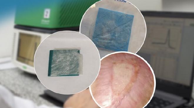
“At AMNIOSBMA, we process the membrane within the first 24 hours after the placenta is delivered. In this process, we sterilize and freeze-dry it, meaning we remove all the moisture. The membrane contains a number of biological factors that begin to degrade after 72 hours. By freeze-drying it, we manage to stop these factors, in a way. Since the patches or dressings we produce are generally used on exudative wounds, applying them rehydrates the membrane with the exudate, generating a sustained release of these beneficial factors. In other words, the moisture from the wound reactivates its properties,” added Mariano Berra, technical director of the NGO.
After 49 days, the area treated with the amniotic membrane dressing showed a much more favorable evolution than the area treated with the animal-derived collagen matrix. “At first glance, it was clear that the skin surrounding the wound had progressed over the ulcer much faster, and the color and type of secretions, in addition to the reduction in inflammatory activity, indicated that the membrane treatment was working better,” explains Guerbi. “Therefore, seeing this macroscopic difference in the evolution of both areas, it was decided to continue the amniotic membrane treatment over the entire wound until it was completely closed,” he added. The entire process of re-epithelialization, or skin repair, lasted approximately five months from the start of treatment.
“In the outermost layers, the maturation of the new tissue was equivalent in both regions, both in the area that originally received the membrane and in the area where it was applied after 49 days. This was expected. Conversely, in deeper layers, maturation was more homogeneous and occurred in less time under the membrane treatment. The correlation between both techniques suggests this improved maturation,” Guerbi pointed out. Furthermore, the team observed that in the area where the animal-derived matrix was applied, a “foreign body” reaction occurred—that is, a rejection of the compound used—and inflammatory processes persisted, something that did not occur in the region where the amniotic membrane was applied. “Vascularization and angiogenesis were also evaluated—that is, how many blood vessels are present and how many new vessels are formed, respectively. An active angiogenesis process was observed, meaning that new vessels began to form under the membrane treatment,” Moreno emphasized.
“When we talk about translational medicine, we’re talking about two worlds that need to connect and engage in dialogue: the world of scientific research steps and methodologies, and the world of medicine, with the complexity of each patient. Here, we were able to translate the knowledge generated in the laboratory, but addressing the patient’s need for an alternative to conventional treatments that hadn’t worked, without resorting to surgery,” Moreno pointed out. Meanwhile, Berra described a virtuous cycle that begins with the reuse of a disposable biological material, with proven qualities for clinical use, and continues on the “front lines,” where doctors and patients encounter specific cases to resolve. “And that, in turn, then returns to the laboratory, where we have to take note of what works and study what factors make it work. All of this is the result of a combination of diverse tasks interacting with each other in an interdisciplinary and fluid way,” she emphasized.
“With the entire regulatory framework approved for human use at the national level, and complying with European regulations for tissue banks, we have been able to bridge the gap that researchers sometimes face when trying to translate scientific developments into clinical applications. In this case, we have already overcome that stage,” emphasized A. Berra, and concluded: “We have demonstrated the potential of amniotic membrane derivatives as a regenerative alternative, and we are now evaluating the mechanism of this process—that is, why and how these regenerative tissues work. We are optimizing a method that in the near future could be performed entirely on an outpatient basis, in the patient’s own home.”
Citation #
-
Guerbi, M. X., Rodrigo, J. M. D. P., Rotela, M. F., Comito, R. A., Vogel, E., Portiansky, E. L., … & Michelini, F. M. (2026). Human Amniotic Membrane Dressing as a Non-Surgical Alternative for Extensive Chronic Ulcers: A Comparative Case Study. International Journal of Molecular Sciences, 27(11), 4655. DOI: https://doi.org/10.3390/ijms27114655
-
Human Amniotic Membrane Dressing as a Non-Surgical Alternative for Extensive Chronic Ulcers: A Comparative Case Study by María Ximena Guerbi, Jimena María del Pilar Rodrigo, Matías Fabián Rotela, Rocío Antonella Comito, Esteban Vogel, Enrique Leo Portiansky, Alejandro Berra, Griselda Noemí Moreno and Flavia Mariana Michelini*, Centro de Medicina Traslacional, Hospital El Cruce (CEMET-HEC), Florencio Varela 1888, Argentina, Comisión de Investigaciones Científicas de la Provincia de Buenos Aires (CIC-PBA), La Plata 1900, Argentina. Instituto de Ciencias de la Salud, Universidad Nacional Arturo Jauretche, Florencio Varela 1888, Argentina. Centro de Educación Médica e Investigaciones Clínicas “Norberto Quirno” (CEMIC), Buenos Aires 1425, Argentina, Servicio de Anatomía Patológica, Hospital El Cruce (HEC), Florencio Varela 1888, Argentina, Laboratorio de Imágenes (CONICET-UNLP), La Plata 1900, Argentina, Banco de Tejidos MNIOS-BMA, San Martín 1650, Argentina, Consejo Nacional de Investigaciones Científicas y Técnicas (CONICET), Buenos Aires 2290, Argentina, Instituto de Estudios Inmunológicos y Fisiopatológicos (IIFP-CONICET), La Plata 1900, Argentina, * Author to whom correspondence should be addressed. Int. J. Mol. Sci. 2026, 27(11), 4655; https://doi.org/10.3390/ijms27114655 Submission received: 1 April 2026 / Revised: 22 April 2026 / Accepted: 24 April 2026 / Published: 22 May 2026 (This article belongs to the Section Molecular Pathology, Diagnostics, and Therapeutics)
Contact [Notaspampeanas](mailto: notaspampeanas@gmail.com)